Breast Imaging
What is the best method of detecting breast cancer as early as possible?
A high-quality mammogram plus a clinical breast exam done by your Women’s Group provider is the most effective way to detect breast cancer early. Finding breast cancer early greatly improves a woman’s chances for successful treatment.
Like any test, mammograms have both benefits and limitations. For example, some cancers can’t be found by a mammogram, but they may be found in a clinical breast exam.
Checking your own breasts for lumps or other changes is called a breast self-exam (BSE). Studies so far have not shown that BSE alone helps reduce the number of deaths from breast cancer. BSE should not take the place of routine clinical breast exams and mammograms. If you notice any unusual changes in your breasts, contact our office to schedule a clinical breast exam.
When should I begin screening mammography?
Everyone has differing medical histories and circumstances. It is best to consult with your physician at your annual exam to recommend a personalized mammography schedule.
Our office follows the general mammogram guidelines from the American Cancer Society (ACS) and the American Congress of Obstetricians and Gynecologists (ACOG):
- Women with an average risk of breast cancer should begin mammograms at age 40 and have them every year.
- Women with a higher risk of breast cancer may benefit by beginning screening mammograms before age 40. Your risk factors and your degree of breast density may lead your doctor to recommend magnetic resonance imaging (MRI) in combination with standard mammograms.
What is my risk for Breast Cancer?
The American Cancer Society’s estimates for breast cancer in the United States for 2014 are:
- About 232,670 new cases of invasive breast cancer will be diagnosed in women.
- About 62,570 new cases of carcinoma in situ (CIS) will be diagnosed (CIS is non-invasive and is the earliest form of breast cancer).
About 5% to 10% of breast cancer cases are thought to be hereditary, meaning that they result directly from gene defects (called mutations) inherited from a parent. Based on your family history of cancers (specifically breast, ovarian, endometrial and colon cancer) you may be in need of diagnostic test that will determine your risk for hereditary cancer syndromes. LINK TO GYNE GENETICS
Please contact our office to schedule a consultation to discuss your cancer risk factors.
Screening mammograms
Screening mammograms are an x-ray exam of the breasts that are used for women who have no breast symptoms or signs of breast cancer or a previous abnormal mammogram. The goal of a screening mammogram is to find breast cancer when it’s too small to be felt by a woman or her doctor. Finding breast cancer early greatly improves a woman’s chance for successful treatment.
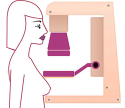
During the mammogram, breasts are compressed between two firm surfaces to spread out breast tissue. A screening mammogram usually takes 2 x-ray pictures of each breast and produces the images on a computer. Some women, such as those with large breasts, may need to have more pictures to see as much breast tissue as possible. This may be uncomfortable, but it’s needed to get a good, clear picture. The pressure only lasts a few seconds.
All mammogram facilities are required to send your results to you within 30 days. In most cases, you will be contacted within 5 working days if there’s a possible problem seen on the mammogram.
How do I get ready for my mammogram?
First, check with the place you are having the mammogram for any special instructions you may need to follow before you go. Here are some general guidelines to follow:
- If you are still having menstrual periods, try to avoid making your mammogram appointment during the week before your period. Your breasts will be less tender and swollen. The mammogram will hurt less and the picture will be better.
- If you have breast implants, be sure to tell your mammography facility that you have them when you make your appointment.
- Wear a two piece outfit, a shirt with shorts, pants, or a skirt. You will be given a gown and asked to undress from the waist up.
- Don’t wear any deodorant, perfume, lotion, or powder under your arms or on your breasts on the day of your mammogram appointment. These things can make shadows show up on your mammogram.
- If you have had mammograms at another facility, have those x-ray films sent to the new facility so that they can be compared to the new films.
- While discomfort from pressure against the breast during the test should be minimal, taking an over-the-counter pain medication such as aspirin, acetaminophen (Tylenol, others) or ibuprofen (Advil, Motrin, others) about an hour before your mammogram can ease any such discomfort.
What can mammograms show?
The radiologist will look at your x-rays for any breast changes that do not look normal and compare these images to your past mammograms. Abnormal findings may include:
- Lump or mass. The size, shape, and edges of a lump sometimes can give doctors information about whether or not it may be cancer. On a mammogram, a growth that is benign often looks smooth and round with a clear, defined edge. Breast cancer often has a jagged outline and an irregular shape.
- Calcifications. A calcification is a deposit of the mineral calcium in the breast tissue. Calcifications appear as small white spots on a mammogram. If calcifications are grouped together in a certain way, it may be a sign of cancer. Depending on how many calcium specks you have, how big they are, and what they look like, your doctor may suggest that you have other tests. Calcium in the diet does not create calcium deposits, or calcifications, in the breast.
What if my screening mammogram shows a problem?
Diagnostic mammogram
A diagnostic mammogram is the next step if an abnormal area is found in a screening mammogram. Diagnostic mammograms are also ordered if a woman has a breast problem (for instance, a lump or nipple discharge). During a diagnostic mammogram, the images are reviewed by the radiologist while you are there so that more pictures can be taken if needed to look more closely at an area of concern. In some cases, special images known as spot views, or magnification views, are used to make a small area of concern easier to evaluate. Often times, a breast ultrasound will be done in addition to the mammogram.
A diagnostic mammogram is usually interpreted in one of three ways:
- It may reveal that an area that looked abnormal on a screening mammogram is actually normal. When this happens, the woman goes back to routine yearly screening.
- It could show that an area of abnormal tissue probably is not cancer, but the radiologist may not be ready to say that the area is normal based on these pictures alone. When this happens it’s common to ask the woman to return to be re-checked, usually in 4 to 6 months.
- The results could also suggest that a biopsy is needed to find out if the abnormal area is cancer. If your doctor recommends a biopsy, it does not mean that you have cancer.
Breast Ultrasound
Ultrasound is an imaging test that uses sound waves to create a picture of your breast. Ultrasound is useful for looking at some breast changes, such as those that can be felt but not seen on a mammogram. It also helps tell the difference between fluid-filled cysts and solid masses. If a lump is really a cyst, it is benign (not cancer).
Magnetic resonance imaging (MRI)
MRI scans use magnets and radio waves instead of x-rays to produce very detailed, cross-sectional pictures of the body. During a breast MRI, a contrast liquid is administered through an IV to help outline the structures of the breast and look for cancer. You will lie on your stomach on a padded platform with spaces for your breasts. You will need to be very still during the test, which can take up to an hour.
Breast MRI is mainly used for 2 purposes:
- For women at high risk for breast cancer, screening MRI is recommended along with a yearly mammogram.
- For women who have been diagnosed with breast cancer, to help measure the size of the cancer and look for any other tumors in the breast. It also can be used to look at the opposite breast to be sure that it does not contain any tumors.
Biopsy
Even though imaging tests like the mammogram and ultrasound can find a suspicious area, they cannot tell whether it’s cancer. A biopsy is the only sure way to diagnose breast cancer.
A biopsy removes some cells from the area of concern so they can be checked under a microscope. The cells can be removed using a needle or by doing surgery to take out part or all of the tumor. The type of biopsy depends on the size and location of the lump or suspicious area.

